Cervical Spondylotic Myelopathy: A Guide to Diagnosis and Management
Neck pain is a nearly universal human experience, often dismissed as a minor consequence of aging or poor posture. However, when degenerative changes in the cervical spine progress to the point of compressing the spinal cord, the condition evolves into Cervical Spondylotic Myelopathy (CSM).
CSM is currently recognized as the most common cause of spinal cord injury in adults, accounting for approximately 54% of non-traumatic spinal cord injuries in North America. Because the onset is often insidious and symptoms can be subtle, diagnosis is frequently delayed—sometimes by more than six years—leading to irreversible neurological damage.
Drawing from a comprehensive analysis of contemporary clinical reviews, systematic meta-analyses, and emerging research into regenerative medicine and machine learning, this article provides an in-depth exploration of the pathophysiology, diagnosis, and management of chronic neck pain and its most severe neurological manifestations.
The Pathophysiology of Cervical Degeneration
The development of CSM is a progressive journey defined by degenerative changes to the vertebrae, intervertebral discs, facets, and ligaments. These changes lead to spinal cord compression through two primary mechanisms: static and dynamic factors.
Static factors are constant mechanical stressors that narrow the spinal canal. These include congenital spinal stenosis, disc herniations, and the development of osteophytes (bone spurs). Over time, ligamentous hypertrophy—specifically the thickening or ossification of the ligamentum flavum or posterior longitudinal ligament—further encroaches upon the canal space. These static obstructions can cause local ischemia (restricted blood flow), leading to neural cell death and apoptosis.
Dynamic factors involve repetitive injury that occurs during normal neck movement. When the neck undergoes flexion or extension, a narrowed canal can cause the spinal cord to stretch or be “squeezed” against compressive lesions.
For example, hyperextension may cause the ligamentum flavum to buckle into the spinal canal, while flexion can stretch axons, making them more susceptible to secondary injury. This cumulative mechanical stress eventually leads to the demyelination of nerve fibers, resulting in the “long-tract signs” that characterize the disease.
Clinical Presentation and the Diagnostic Challenge
The hallmark of CSM is its subtle, often “benign” beginning. Patients rarely present with sudden paralysis; instead, they report a gradual decline in function.
Initial Symptoms and “Myelopathy Hand”
The most frequent initial complaints involve decreased hand dexterity and gait instability. Patients may find it difficult to perform fine motor tasks such as buttoning a shirt, handwriting, or typing. Clinical examinations often reveal “myelopathy hand,” characterized by a positive “finger escape sign” (an inability to keep the small fingers adducted) or a deficient “grip and release test” (an inability to rapidly make and open a fist).
Gait and Lower Extremity Involvement
As the disease progresses, gait dysfunction becomes the hallmark sign, affecting over 80% of patients. This is often characterized by a wider step width, shorter step length, and a general feeling of instability or frequent falls. Physicians may observe upper motor neuron signs in the lower extremities, including hyperreflexia, a positive Babinski reflex (which is 100% specific for CSM), and sustained foot clonus.
Special Physical Exams
Several specific maneuvers can aid in the diagnosis:
- Hoffman Sign: Involuntary flexion of the thumb or index finger when the middle finger nail is flicked. While it has a 68% positive predictive value, it remains a useful adjunct in a broader clinical workup.
- Hyperactive Pectoralis Reflex: A highly sensitive (84.8%) and specific (96.7%) marker for cord compression at the C2 to C4 levels.
- Lhermitte’s Phenomenon: An electric shock-like sensation that runs down the spine or into the limbs upon neck flexion, reported by approximately 27% of patients.
The Future of Diagnosis: Machine Learning and AI
One of the most innovative frontiers in managing neck pain is the use of discomfort drawings coupled with machine learning. Traditionally, patients shade areas of pain on a body contour to help clinicians visualize symptoms. Recent research has explored the Inter-Battery Topic Model (IBTM), a generative machine learning framework that can predict diagnostic labels—such as radiculopathy or disc-ligament injury—based solely on these drawings.
By treating a patient’s discomfort drawing as a “bag-of-location words,” AI models have shown reasonable success in discerning neuropathic pain from other types of discomfort. In experimental settings, these models have achieved high interpretive accuracy, suggesting that AI could soon serve as a vital decision-support system for primary care physicians, helping to flag potential CSM cases earlier in the disease’s progression.
Radiographic Evaluation: The Gold Standard
While physical exams provide critical clues, imaging is required to confirm the diagnosis and plan treatment.
- Magnetic Resonance Imaging (MRI): The gold standard for CSM evaluation. MRI allows for the visualization of soft tissues, disc herniations, and intrinsic cord abnormalities, such as T2 signal intensity changes, which indicate cord edema or myelomalacia.
- Radiographs (X-rays): Still useful for assessing spinal alignment and calculating the Torg-Pavlov ratio. A ratio of less than 0.8 signifies canal stenosis, while a canal diameter of less than 12mm is highly correlated with cord compression.
- Computed Tomography (CT) Myelography: Primarily used for patients with contraindications to MRI. It remains the “gold standard” for diagnosing the ossification of the posterior longitudinal ligament.
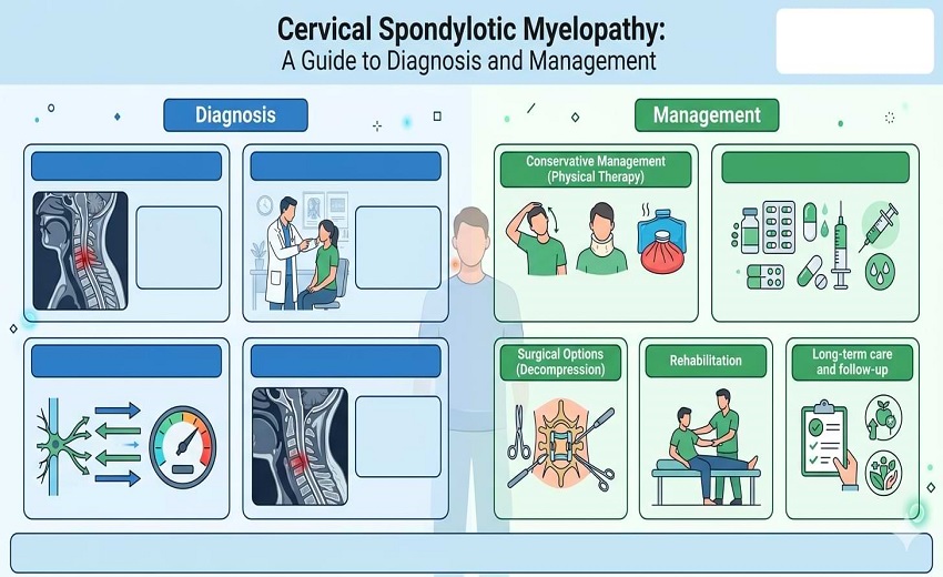
Management Strategies: Operative vs. Non-Operative
The management of CSM is dictated by the severity of the symptoms, typically graded using the Japanese Orthopaedic Association (JOA) scale.
Non-Operative Care
For patients with mild CSM (JOA score 15–17), a supervised trial of structured rehabilitation may be offered. Conservative treatments include medications, thermal therapy, and soft collars. However, clinicians are cautioned against spinal manipulation or heavy traction, as anecdotal reports suggest these may worsen neurological symptoms in myelopathic patients.
Surgical Intervention
CSM is generally considered a surgical disease because non-operative management often fails to prevent progression; up to 56% of patients managed non-operatively experience significant impairment within 10 years.
- Anterior Approaches: Procedures like Anterior Cervical Discectomy and Fusion (ACDF) or Corpectomy (ACCF) are preferred for patients with kyphosis or 1-2 level involvement.
- Posterior Approaches: Laminectomy (with or without fusion) and laminoplasty are typically reserved for multi-level disease or congenital stenosis.
A recent systematic review of over 22,000 patients revealed that while ACDF provides high fusion rates, it may be associated with higher complication rates compared to less invasive posterior foraminotomies or laminoplasty. Complications can include dysphagia (difficulty swallowing), C5 palsy, and pseudarthrosis (failure of the bone to fuse).
Regenerative Medicine: The Next Decade
The most promising shift in cervical spine care is the emergence of regenerative medicine and stem cell therapy. Research into Mesenchymal Stem Cells (MSCs)—sourced from bone marrow or umbilical cords—has shown the potential to move treatment from “symptom control” to “tissue restoration”.
Experimental applications currently include injecting stem cells directly into degenerated discs to modulate the immune environment and promote the regeneration of the extracellular matrix. Early clinical trials have reported significant pain reduction and improved range of motion in patients with discogenic pain.
Furthermore, using MSCs as an adjuvant to surgery has been shown to increase fusion rates and patient satisfaction in ACDF procedures compared to bone grafts alone. While these therapies remain largely investigational and are not yet “standard of care,” they represent a potential revolution in how we treat the aging spine.
Conclusion and Referral Guidelines
The primary challenge in managing neck pain is the “asymptomatic” nature of early cord compression. Up to 31% of the population over age 50 has MRI evidence of cord compression, yet only a fraction will develop clinical myelopathy.
For the primary care provider, the directive is clear: early referral is essential. Because advanced age, long duration of symptoms, and severe preoperative deficits are the strongest predictors of poor surgical outcomes, the window for effective intervention is narrow. Any patient presenting with “clumsy hands,” gait instability, or hyperreflexia should be considered for urgent specialist evaluation.
As we integrate AI-driven diagnostic tools and regenerative therapies into clinical practice, the goal remains to identify these “insidious” changes before they result in permanent disability, transforming the management of neck pain from reactive treatment to proactive restoration.